Disposable titanium-nickel memory alloy fistula stapler
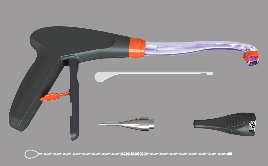
- Disposable titanium-nickel memory alloy fistula stapler
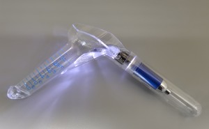
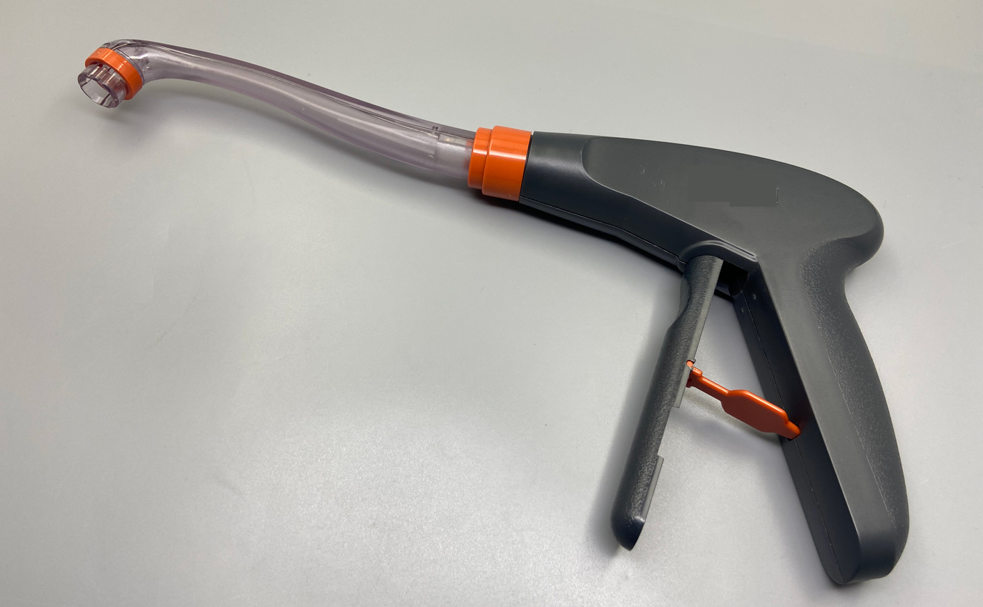
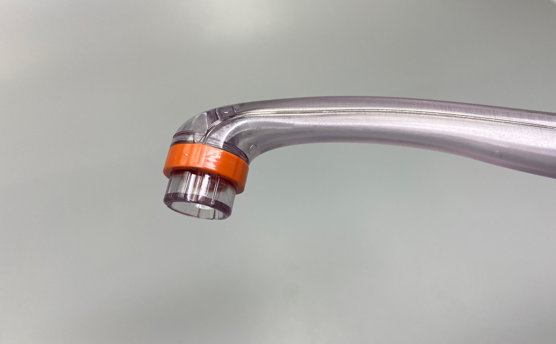
- fistula stapler
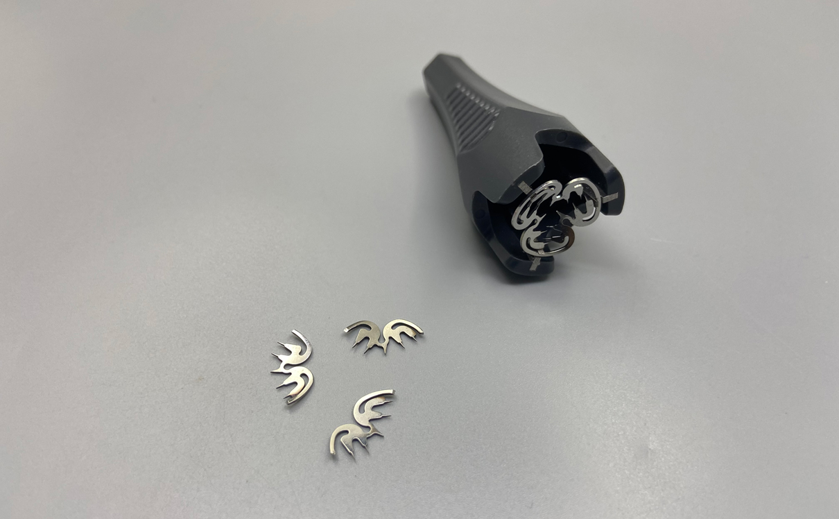
- fistula stapler pusher
- Fistula stapler push sleeve and stapler clip
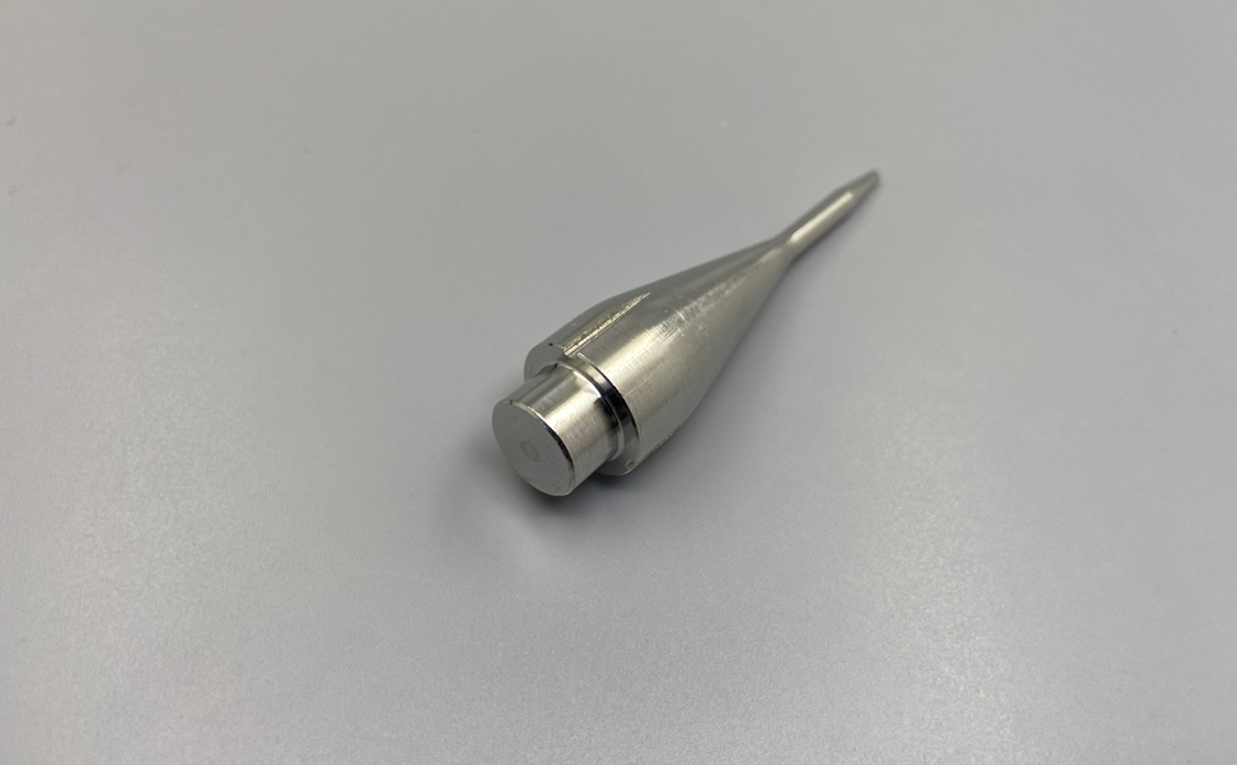
- fistula stapler guide
- Fistula Stapler Fistula Brush

Instructions:
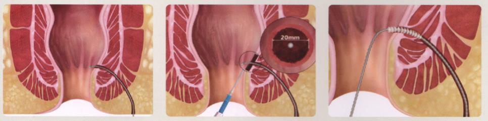
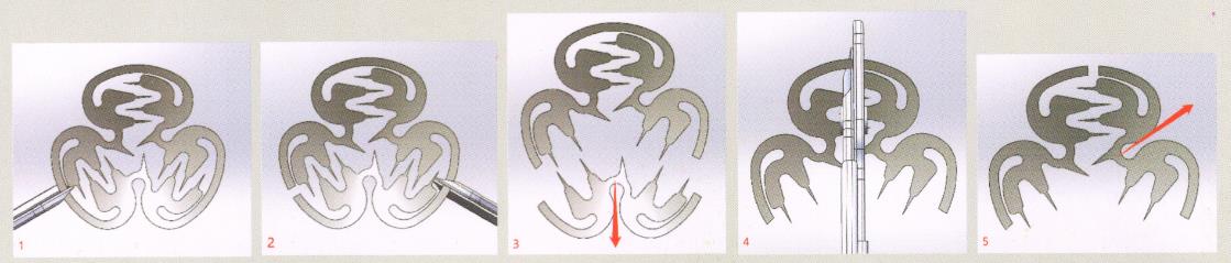
Figure 1: Probing the internal opening with a probe.
Figure 2: The mucosal tissue with a diameter of 1.5 cm to 2 cm around the internal opening was excised with an electric knife.
Figure 3: Use an anal fistula brush or other means to clear the fistula tract.
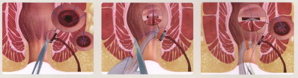
Figure 4: Purse-string closure of the internal opening with 3-0 absorbable sutures.
Figure 5 and Figure 6: Use the suture to pull the tissue around the internal opening into the cavity of the head of the dispenser, and release the anastomotic clip to complete the clipping.
Precautions
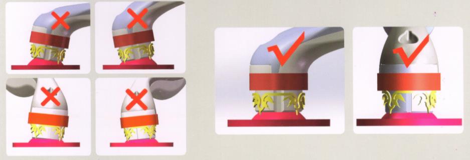
Releasing the anastomotic clip is a key step in the operation. It should be ensured that enough muscle tissue is pulled in before release. The strength of pulling the muscle tissue should not be relaxed during release, and the anastomotic clip should be released vertically. The anastomotic clips should all be clipped in the muscular layer and not brought into the mucosal tissue. Improper release may result in premature dislodgement of the anastomotic clip, resulting in surgical failure.
The anastomotic clip is designed to be detachable. Usually, it can fall off on its own at about 3 weeks after the operation and be excreted with the stool. If it does not fall off at 4 weeks, it should be removed manually in order to avoid being embedded by the mucous membrane and forming foreign bodies in the body.