
Functional characteristics of laparoscopic trainer
The training manikin of laparoscopic surgery skills can be used for the simulation training of laparoscopic surgery for common abdominal diseases with laparoscopic surgical instruments, high-definition cameras and monitors on the operating table in surgery, gynecology and obstetrics. It can carry out the basic operations of laparoscopic surgery, such as incision, stripping, hemostasis, ligation, suture and so on.
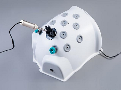
The simulated laparoscopic 30 degree mirror can achieve the purpose of multi-directional observation. The light source LED and camera are embedded in the lens. The field of vision image in the abdominal cavity of the manikin is output to the 22 inch color screen, and the operator operates by observing the image on the screen.
The simulated laparoscope can adjust the focal length by stretching and adjusting the distance between the lens and the target to change the clarity of the image. When the lens is close to the intra-abdominal model, it can obtain a locally enlarged image, and when it retreats to the opening of the cannula, it can obtain a wider field of vision in the abdominal cavity. It can be adjusted in time according to the precision of operation and observation needs. The central field of vision of the lens should move with the instrument of the prospective operator, and adjust the short-range or long-range field of vision as needed.
Various training models can be placed in the simulated abdominal cavity, including: colored bean model, ferrule model,suture plate model, multi shape suture model, cystic organ model, cecal appendix model, liver and gallbladder model, uterus and accessories model, threading model, transverse colon model, kidney and ureter model, pancreas and spleen model, vascular model, intestinal model, organ adhesion model. One of various training models can be selected according to teaching needs, Put it into the abdominal cavity.
Ferrule model: Six inverted L-shaped steel hooks are set on the cylindrical rubber block, and the trainees use claws to grasp the small loop and put it on it until it is full. Repeated training can gradually improve the speed.
Colored bean model: grab the colored beans of various colors in the container, grab the specified colors, and grab them in their respective containers.
Threading model: the top of more than 10 conical rubber blocks is equipped with a steel ring with a diameter of 2-3mm. The suture is clamped with a needle holder and passed through the steel ring one by one until the threading is completed.
Cystic organ model: the thin part can be cut and anastomosed, and the swollen part can be cut and sutured or cut and anastomosed.
Vascular model: small vessel ligation training can be carried out.
Models of various internal organs: when used, they are pasted on the back plate to prevent movement during operation. Various organs can be cut, stopped bleeding, stripped, sutured and knotted.
Liver gallbladder model: cholecystectomy training can be carried out.
Kidney and ureter model: ureteral anastomosis and stone removal can be performed.
Intestinal model: intestinal (incision) anastomosis can be performed.
Cecal appendix model: Appendectomy training can be carried out, other organs can be practiced such as stripping, resection and suture, and the simulated appendiceal artery and gallbladder artery can be replaced.
Training on operation skills of simulated laparoscopic trainer
Through training, beginners of abdominal malocclusion surgery can begin to adapt to the transition from stereovision under direct vision to plane vision of the monitor, carry out orientation and coordination adaptation, and choose various instrument operation skills.
There are not only differences in depth, size, but also differences in vision, orientation and movement coordination between laparoscopic surgery and direct vision surgery. Beginners must be trained to adapt to this change. One of the conveniences of direct vision surgery is that the stereovision formed by the operator's two eyes can distinguish the position between far and near and between each other due to different visual angles when observing objects and surgical fields, and carry out accurate manipulation. The images obtained by laparoscopy, camera and television monitoring system are quite dry from monocular vision and lack stereoscopic sense, so it is easy to produce errors when judging the distance between far and near. To the color eye effect formed by the dry endoscope (when the abdominal cavity is slightly deflected, the same object will show different geometric shapes on the TV screen), the operator must gradually adapt. Therefore, in the training, we should learn to grasp the size of each object in the image, estimate the distance between them and the wrong plane of the abdominal staggered objective in combination with the size of the original entity, and operate the instrument. The operator and assistant should consciously strengthen the sense of plane vision, and judge the exact position of the instruments and organs according to the shape and size of the organs and instruments at the surgical site after light microscopy, and the intensity of the image light.
Normal orientation and coordination ability are the necessary conditions for successful operation. The operator determines the target orientation and distance according to the information obtained from vision and orientation, and the motion system coordinates the action to operate. This has formed a complete reflection in daily life and direct vision surgery, and is used to it. Endoscopic operation, such as cystoscopic ureteral intubation, is easy to adapt to the orientation and movement coordination of the operator because the direction of the short mirror is consistent with the direction of operation. However, when the TV abdominal surgery is wrong, the orientation and coordination formed by previous experience often lead to wrong operation, such as the operator standing on the left side of the supine patient, and the TV screen is not placed on the foot of the patient. At this time, the TV image shows the position of Jing Yi, The operator will habitually extend the instrument to the direction of the TV screen, and mistakenly believe that this is approaching Jingyi, but in fact, the instrument should be extended to the deep surface to reach the seminal vesicle. This is the directional reflection formed by direct vision surgery and wrong endoscope operation in the past. When the TV abdominal surgery is wrong, it will not work. When observing the TV image, the operator should consciously determine the relative position between the instrument in his hand and the relevant organs in the patient's abdomen, and make appropriate advance and retreat, Only by rotating or tilting, and mastering the amplitude, can the exact clamp be carried out at the surgical site. The operator and assistant should determine the orientation of their instruments from the same TV image according to their respective positions before they can cooperate with the operation. The position of the laparoscope should be changed as little as possible. A little rotation may rotate or even reverse the image, making orientation and coordination more difficult. Practicing in the training box or oxygen bag for many times and cooperating with each other can make the orientation and coordination ability better adapt to the new situation, shorten the operation time and reduce trauma.
Post time: Jul-08-2022


