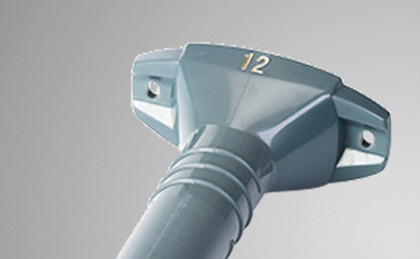
Thoracic indwelling tube - closed thoracic drainage
1 Indications
1. A large number of pneumothorax, open pneumothorax, tension pneumothorax, pneumothorax oppresses the breathing (generally when the lung compression of unilateral pneumothorax is more than 50%).
2. Thoracocentesis in the treatment of lower pneumothorax
3. Pneumothorax and hemopneumothorax requiring mechanical or artificial ventilation
4. Recurrent pneumothorax or hemopneumothorax after removal of thoracic drainage tube
5. Traumatic hemopneumothorax affecting respiratory and circulatory functions.
2 Preparation
1. Postures
Sitting or semi reclining position
The patient is in the half lying position (if the vital signs are not stable, the patient is in the flat lying position).
2. Pick site
1) Selection of the second intercostal space of the middle clavicular line for pneumothorax drainage
2) Pleural effusion was selected between the axillary midline and the posterior axillary line, and between the 6th and 7th intercostals
3. Disinfection
Routine skin disinfection, diameter 15, 3 iodine 3 alcohol
4. Local infiltration anesthesia
Intramuscular injection of phenobarbital sodium 0 lg。
Local infiltration of the chest wall preparation layer in the anesthesia incision area to the pleura; Cut the skin 2cm along the intercostal line, extend the vascular forceps along the upper edge of the ribs, and separate the intercostal muscle layers to the chest; The drainage tube shall be placed immediately when liquid gushes out. The depth of the drainage tube into the chest cavity should not exceed 4 ~ 5cm. The chest wall skin incision should be sutured with medium-sized silk thread, the drainage tube should be ligated and fixed, and covered with sterile gauze; Outside the gauze, wrap a long tape around the drainage tube and paste it on the chest wall. The end of the drainage tube is connected to the disinfection long rubber tube to the water sealed bottle, and the rubber tube connected to the water sealed bottle is fixed on the bed surface with adhesive tape. The drainage bottle is placed under the hospital bed where it is not easy to be knocked down.

3 Intubation
1. Incision of skin
2. Blunt separation of muscular layer and placement of thoracic drainage tube with side hole through the upper edge of rib
3. The side hole of the drainage tube should be 2-3cm deep into the chest cavity
4 Precautions
1. In case of massive hematocele (or effusion), the blood pressure should be closely monitored during the initial drainage to prevent the patient from sudden shock or collapse. If necessary, the blood pressure should be continuously released to avoid sudden danger.
2. Pay attention to keep the drainage tube unblocked without pressure or distortion.
3. Help the patient to change the position properly every day, or encourage the patient to take a deep breath to achieve full drainage.
4. Record the daily drainage volume (the drainage volume per hour in the early stage after injury) and the changes of its properties, and conduct X-ray fluoroscopy or film reexamination as appropriate.
5. When replacing the sterile water sealed bottle, the drainage tube shall be temporarily blocked first, and then the drainage tube shall be released again after the replacement to prevent the air from being sucked in by the negative pressure of the chest.
6. In order to eliminate secondary infection, bacterial culture and drug sensitivity test of drainage fluid can be performed if the drainage fluid properties are changed.
7. When pulling out the drainage tube, the skin around the incision should be disinfected first, the fixed suture should be removed, the drainage tube near the chest wall should be clamped with vascular forceps, and the drainage opening should be covered with 12 ~ 16 layers of gauze and 2 layers of Vaseline gauze (including a little more vaseline is preferred). The operator should hold the gauze with one hand, hold the drainage tube with the other hand, and pull it out quickly. The gauze at the drainage opening was completely sealed on the chest wall with a large piece of adhesive tape whose area exceeded the gauze, and the dressing could be changed after 48 ~ 72 hours.
5 Postoperative nursing
After operation, the drainage tube is often overstocked to keep the lumen unobstructed. The drainage flow is recorded every hour or 24 hours. After drainage, the lung expands well, and there is no gas or liquid outflow. The drainage tube can be removed when the patient inhales deeply, and the wound can be closed with vaseline gauze and adhesive tape.
Post time: Jun-10-2022


