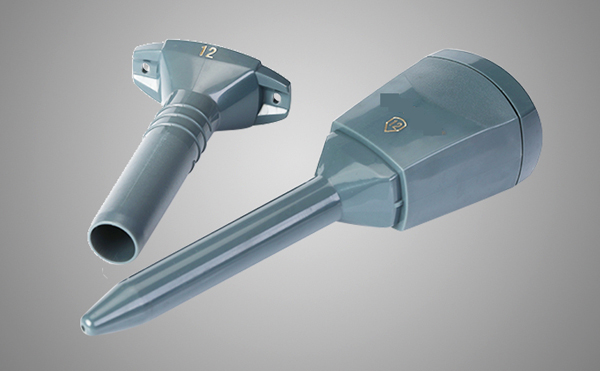
Thoracentesis
1、 Indications
1. Pleural effusion of unknown nature, puncture test
2. Pleural effusion or pneumothorax with compression symptoms
3. Empyema or malignant pleural effusion, intrapleural administration
2、 Contraindications
1. Uncooperative patients;
2. Uncorrected coagulation disease;
3. Respiratory insufficiency or instability (unless relieved by therapeutic thoracentesis);
4. Cardiac hemodynamic instability or arrhythmia; Unstable angina pectoris.
5. Relative contraindications include mechanical ventilation and bullous lung disease.
6. Local infection must be excluded before the needle penetrates the chest.
3、 Complications
1. Pneumothorax: pneumothorax caused by gas leakage of puncture needle or lung trauma under it;
2. Hemothorax: pleural cavity or chest wall hemorrhage caused by puncture needle damaging subcostal vessels;
3. Extravasated effusion at puncture point
4. Vasovagal syncope or simple syncope;
5. Air embolism (rare but catastrophic);
6. Infection;
7. Stabbing injury of spleen or liver caused by too low or too deep injection;
8. Relapsing pulmonary edema caused by rapid drainage > 1L. Death is extremely rare.

4、 Preparation
1. Postures
In the sitting or semi reclining position, the affected side is on the side, and the arm of the affected side is raised above the head, so that the intercostals are relatively open.
2. Determine the puncture point
1) Pneumothorax in the second intercostal space of the middle clavicular line or 4-5 intercostal spaces of the middle axillary line
2) Preferably the scapular line or the 7th to 8th intercostal space of the posterior axillary line
3) If necessary, 6-7 intercostals of axillary midline can also be selected
Or the 5th intercostal space of axillary front
Outside the costal angle, blood vessels and nerves run in the costal sulcus and are divided into upper and lower branches at the posterior axillary line. The upper branch is in the costal sulcus and the lower branch is at the upper edge of the lower rib. Therefore, in thoracocentesis, the posterior wall passes through the intercostal space, close to the upper edge of the inferior rib; The anterior and lateral walls pass through the intercostal space and through the middle of the two ribs, which can avoid damaging intercostal vessels and nerves.
The positional relationship between blood vessels and nerves is: veins, arteries and nerves from top to bottom.
The puncture needle should be inserted into the intercostal space with liquid. There is no encapsulated pleural effusion. The puncture point is usually a costal space below the liquid level, located at the infrascapular line. After the skin was disinfected with iodine tincture, the operator wore sterile gloves and laid a sterile hole towel, and then used 1% or 2% lidocaine for local anesthesia. First make a colliculus on the skin, then subcutaneous tissue, periosteum infiltration on the upper edge of the lower rib (to prevent contact with the lower edge of the upper rib to avoid damaging the subcostal nerve and vascular plexus), and finally to the parietal pleura. When entering the parietal pleura, the anesthesia needle tube can suck the pleural fluid, and then clamp the anesthesia needle with a vascular clamp at the skin level to mark the depth of the needle. Connect the large caliber (No. 16~19) thoracentesis needle or needle cannula device to a three-way switch, and connect a 30~50ml syringe and a pipe to empty the liquid in the syringe into the container. The doctor should pay attention to the mark on the anesthesia needle that reaches the chest fluid depth, and then inject the needle for 0.5cm. At this time, the large-diameter needle can enter the chest cavity to reduce the risk of penetrating the underlying lung tissue. The puncture needle vertically enters the chest wall, subcutaneous tissue, and enters the pleural fluid along the upper edge of the lower rib. The flexible catheter is superior to the traditional simple thoracentesis needle because it can reduce the risk of pneumothorax. Most hospitals have disposable chest puncture discs designed for safe and effective puncture, including needles, syringes, switches and test tubes.
Post time: Jun-06-2022


